One of the most important requirements for creating accurate 3D skeletal models is being able to generate high-quality digitised bones. For our project we are using two methods: computed tomography (CT) scanning, and photogrammetry.
Computed Tomography (CT scanning)
CT scanning is probably the most widely known method used in palaeontology for obtaining digital fossils as it is regularly used in medicine for looking at skeletons and bones to diagnose illnesses. CT scans are created by taking series of x-rays of an object from different orientations (usually circles or helically) and from those extracting the 3D views of the x-rays which we call CT scans. From these CT scans we are able to use computer software to highlight the objects of specific interest (a process called segmentation). CT scanning is an incredibly powerful tool for palaeontology as it allows us to segment fossil bones that are still encased in the rock, as well as getting internal anatomy of the bones that otherwise would not be possible from looking at the surfaces.
The image below shows segmented CT scan data of the right hindlimb (on left) and forelimb (on right) of a young Nile crocodile; in top view; used in our studies. To enable modelling of biomechanics, each bone to be handled independently is coloured differently in segmentation.

Photogrammetry
Photogrammetry is a rapidly growing source for digital models as they can be created using just a camera (even a mobile phone camera will work), and freely available software (unlike CT scans which involve expensive machinery with x-rays). Photogrammetry works by capturing photographs of an object from a series of orientations and software stitches together the photographs using identical points (either picked manually or automatically) to create what is functionally a 3D panoramic of the object. The more photographs and orientations that there are, the more points of similarity can be found by the software, and the higher the accuracy of the model. This method captures surface data and lacks the internal structure of bones, but this is not necessary for all models. It also allows the capture of mounted (exhibited) fossils that cannot be taken off display.
Mass Modelling
Once a skeleton has been imaged in 3D, computer graphics software can be used to reconstruct the fleshy boundaries of the whole body and its individual segments (head, thigh, tail, etc.). Segments can then be given densities and thereby volumes can be used to calculate masses, centres of mass and inertia of those segments and the whole body. Such data are critical for estimating how extinct archosaurs stood and moved (e.g. our published study of Mussaurus); and also for simulating motion in extant archosaurs. This procedure has been fairly well-honed for archosaurs by our and others’ teams.
The images below show the mass modelling technique used in the DAWNDINOS project for Batrachotomus.
a) Photogrammetry software is used to construct a digital skeleton from photos,
b) The digital skeleton is placed in a standardised pose,
c) Volumes representing flesh and airspaces are added to the skeleton,
d) Finished volumetric model,
e) Estimates of body mass, inertia and centre of mass calculated from body volume.
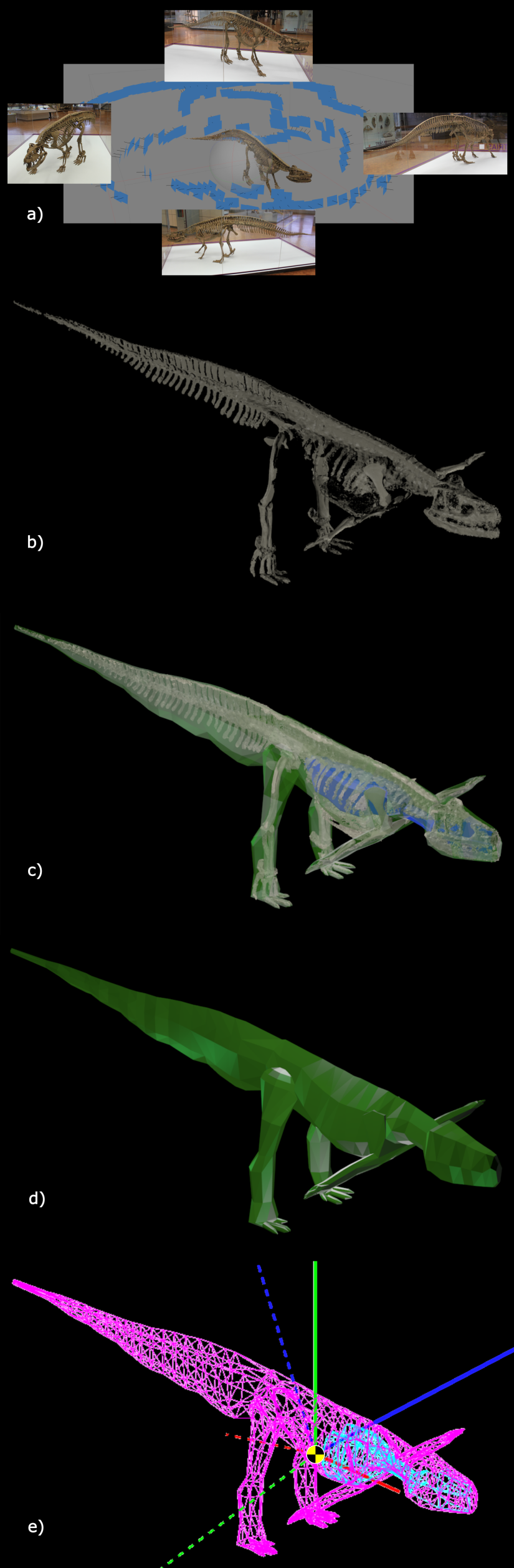
Mass modelling of Batrachotomus
